Advanced Imaging
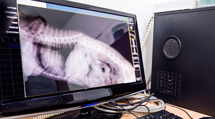
Here at Oakview, we pride ourselves on using the most advanced imaging techniques in order to help your pets faster and more efficiently. Therefore, we have digital x-rays, which produce a much higher quality image and can be placed on CDs or emailed to specialists.
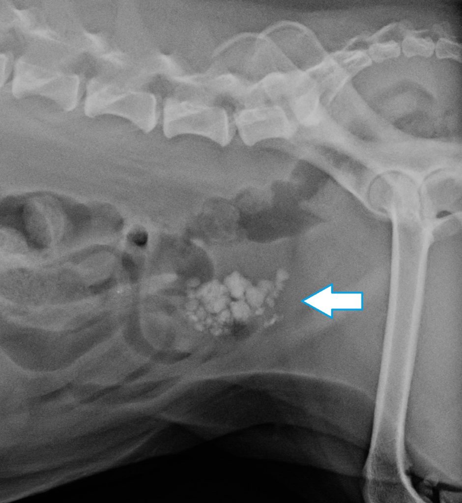
Here is an example of a digital x-ray. The darker areas are gas in the intestines. The blue arrow is pointing to a grouping of bright white spots. These are bladder stones.
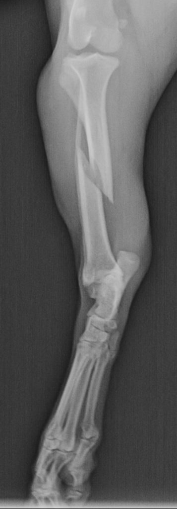
This is a digital x-ray of a dog’s rear leg. Can you find the fracture?
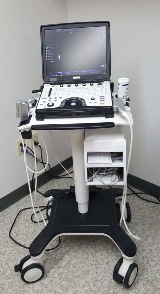
We also have an ultrasound machine which enables us to visualize soft tissue better than x-rays.
Dr. Valerie Curtis is our sonographer. She has a special interest in ultrasound and has enhanced her knowledge through continuing education.
- To evaluate masses in the abdomen (although only an actual biopsy can diagnose)
- To evaluate liver problems revealed in blood work
- To enable sterile urine collection
- To characterize fluid in the chest or abdomen
- To do a pregnancy check (surprisingly, we cannot count the babies this way, they are too mobile!)
Why does my pet’s hair need to be shaved for an ultrasound?
The ultrasound probe needs to be in direct contact with the skin in order to transmit readable images.
Why might my pet need sedation for an ultrasound?
In order to gather clear pictures, the pet cannot move. It would be like trying to take a still picture of a moving target–only blurry images result.
Our dental x-rays are digital and help evaluate your pet’s teeth, jaw, and gum issues.
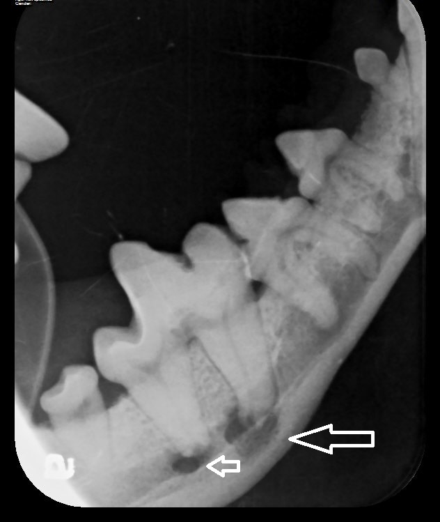
This dog came in for a routine dental cleaning. He had no symptoms of pain. However, this dental x-ray revealed a painful tooth root abscess! This is reflected by the dark pockets at the base of the tooth. Pets don’t always show us their pain, so advanced imaging is one tool we can use to find it.
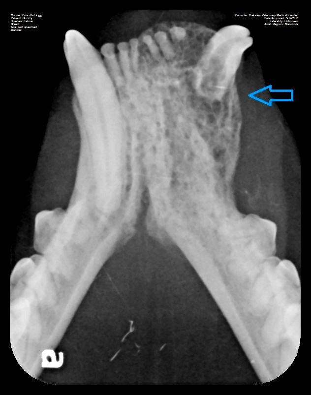
This digital dental x-ray helped to evaluate a growth in the mouth shown by the blue arrow.
Penn Hip Evaluations
Hip Dysplasia and Arthritis are the major orthopedic conditions that we see in dogs. The majority of the time there is no cure and is treated with medical management. Early detection is key to this. At the , we now offer Penn Hip evaluation. This is a diagnostic tool that looks at the laxity or looseness of the hips of your pet. This determination is done by taking 3 radiographs under anesthesia. Based on the views your dog will receive a Distraction Index (DI). The DI ranges from 0 to 1. Those pets closer to 1 mean there is more laxity/looseness and more prone to developing hip dysplasia. Evaluation of a dog’s hip can be done as soon as 16 weeks.
We normally recommended this evaluation be done at the time of spay or neuter to eliminate the need for extra anesthetic procedures. However, in some puppies who are experiencing pain in the hind end and/or are high-risk breeds, it may be beneficial to perform sooner. This is because there is a procedure that could be done to prevent the progression of the disease, but it needs to occur before pets reach full maturity. High-risk breeds include Golden Retrievers, Labradors, German Shepherds, and Saint Bernards, however, all large breeds can be susceptible.
The evaluation can be imperative to breeders as well. The lower the DI, the less chance of offspring developing hip dysplasia. Please call the clinic to schedule with our certified doctor, Dr. Lauren, or if you have any questions.
OFA Radiographs
To ensure your pet has healthy joints in their elbows and hips, our doctors can perform xrays under the Orthopedic Foundation for Animals guidance. These images can help rule out hip dysplasia and elbow dysplasia. To learn more, please give us a call at 715-344-6311.
Advanced Imaging Animal Hospital Plover, Wisconsin
Visit Oakview Veterinary Medical Center for more information on our animal advanced imaging. Fill out our contact form or give our hospital a call at 717-344-6311 to schedule an appointment.
